 EGCG (epigallocatechin gallate) is the most abundant and perhaps most important catechin found in green tea (camellia sinensis). Catechins are polyphenolic antioxidant plant metabolites and belong in the flavonoid family. EGCG functions as a powerful antioxidant, preventing oxidative damage in healthy cells, but also as an antiangiogenic and antitumor agent and as a modulator of tumor cell response to chemotherapy.
There is a huge literature showing that EGCG kills cancer cells of all kinds. EGCG reactivates epigenetically silenced genes in cancer cells and induces apoptosis and promotes cell growth arrest, by altering the expression of cell cycle regulatory proteins, activating killer caspases, and suppressing nuclear factor kappa-B (NF-kB) activation.
The majority of human cancers demonstrate the inactivation of the p53 pathway. p53 is one of the most frequently mutated tumor suppressor genes in human cancers. EGCG increases p53 transcriptional activity and acetylation by suppressing class I HDACs (histone deacetylases), a function that is likely to be part of the mechanisms that control the physiological activity of p53. Acetylation of p53 at the carboxy-terminal lysine (Lys) residues enhances its transcriptional activity associated with cell cycle arrest and apoptosis.
EGCG-induced stabilization of p53 protein resulted in the regulation of p21 and Bax, thereby positively changing the ratio of Bax/Bcl-2, activating initiator and effector caspases and PARP cleavage, leading to the induction of apoptosis. Besides, EGCG regulates and promotes IL-23 dependent DNA repair and stimulates cytotoxic T cells activities in a tumor microenvironment.
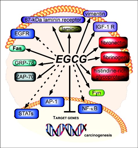
EGCG has been reported to directly bind with the plasma proteins fibronectin, fibrinogen and histidine-rich glycoprotein, Fas, laminin and the 67 kDa laminin receptor, vimentin ZAP-70, Fyn, insulin-like growth factor-I receptor (IGF-IR), and glucose-regulated protein 78 (GRP-78). EGCG also indirectly targets a number of other oncogenic proteins including EGFR, AP-1, and STAT. It also blocks carcinogenesis by modulating the signal transduction pathways, including JAK/STAT, MAPK, PI3K/AKT, Wnt and Notch, involved in cell proliferation, transformation, inflammation and metastasis.
Epigallocatechin 3-gallate and green tea catechins: United they work, divided they fail.
Modulation of signaling pathways in prostate cancer by green tea polyphenols.
EGCG inhibits growth, invasion, angiogenesis and metastasis of pancreatic cancer.
Furthermore, EGCG stimulates telomere fragmentation through inhibiting telomerase activity, thereby increasing cellular apoptosis and inhibiting cellular proliferation. When cell divide, the ends of the DNA, called telomeres, shorten after every cell division. Eventually, this leads to the death of the cell. Pure and simple, this is a fundamental aspect of normal cell aging.
Unlike normal cells, cancer cells can grow and age without dying. The enzyme telemerase repairs the damaged DNA, thereby increasing the lifespan of the cells. Normal cells contain very low levels of telemerase. The normal cells will divide for a number of divisions and then stop growing. They get old and either they die or they sit there and do nothing. They are mortal. Cancer cells, on the other hand, all cancer cells, contain very high levels of this enzyme.
Cancer cells have very short telomeres. Their telomeres don’t get shortened. This allows cancer cells to escape one of the major aging controls. The only thing keeping these cells alive is their over expression of the enzyme telomerase. Once the ends of the DNA shorten to a prescribed length, the cells will die by programmed cell death. So inhibition of telomerase activity is a major goal of the natural cancer treatment.
Pharmaceutical regulation of telomerase and its clinical potential.
Epigenetic and genetic mechanisms contribute to telomerase inhibition by EGCG.
EGCG, on the other hand, is a powerful natural proteasome inhibitor at very low doses. The proteasome is a multicatalytic proteinase complex responsible for the degradation of most intracellular proteins, including proteins crucial to cell cycle regulation and programmed cell death, or apoptosis. Targeting the proteasome has become an attractive approach in cancer therapy.
Structure-activity study of epi-gallocatechin gallate (EGCG) analogs as proteasome inhibitors.
Green tea polyphenols as a natural tumour cell proteasome inhibitor.
Green tea polyphenols as proteasome inhibitors: implication in chemoprevention.
Ester bond-containing tea polyphenols potently inhibit proteasome activity in vitro and in vivo.
Targeting tumor ubiquitin-proteasome pathway with polyphenols for chemosensitization.
Proteasome inhibitors are much more toxic to leukemia and lymphoma cells than they are to sarcoma and carcinoma cells. The inhibition of the proteasome complex which is absolutely necessary for immune functioning is an excellent way to kill B and T cell lymphomas and leukemias. Immune cells all depend on the activation of NF-kB (a proinflammatory transcription factor) for growth and survival. But, sarcoma and carcinoma cells are less susceptible to the inhibition of NF-kB signaling.
Proteasome inhibitors are known to decrease the level of activated NF-kB in cells. Since the inhibition of the proteasome blocks the activation of NF-kB, proteasome inhibitors such as EGCG would have a stronger therapeutic affect against lymphomas and leulemias than sarcomas and carcinomas.
Velcade (Bortezomib) represents the first proteasome inhibitor to have shown anti-tumor activity in both solid and haematological malignancies. One of the major mechanisms of Velcade is through upregulation of NOXA, which is a proapoptotic protein, and NOXA may interact with the anti-apoptotic proteins of Bcl-2 subfamily Bcl-X(L) and Bcl-2, and result in apoptotic cell death in malignant cells.
Another important mechanism of Velcade is through suppression of the NF-κB signaling pathway, resulting in increased apoptosis, decreased angiogenic cytokine production, and inhibition of tumor cell adhesion to stroma. Moreover, Velcade, mainly by inhibition of the NF-kB pathway, has a chemosensitizing effect when administered together with other antitumoral drugs.
The potential of proteasome inhibitors in cancer therapy.
Proteasome inhibition suppresses essential immune functions of human CD4+ T cells.
Bortezomib in the treatment of cancer.
Advances in the understanding of mechanisms and therapeutic use of bortezomib.
Current status of bortezomib in the treatment of multiple myeloma.
NF-kappaB as a therapeutic target in chronic lymphocytic leukemia.
Although chemotherapy is a therapeutic strategy for cancer treatment, it fails to eliminate all tumor cells due to intrinsic or acquired drug resistance, which is the most common cause of tumor recurrence. Furthermore, NF-kB is induced by all chemotherapy agents and certainly all doses of radiation resulting in a loss of therapeutic effectiveness by these agents.
EGCG has exhibited an appreciable effect on overcoming resistance to various chemotherapeutic drugs as well as multidrug resistance (MDR) in a broad spectrum of tumors ranging from carcinoma and sarcoma to hematological malignances. EGCG acts to enhance the cancer killing properties of the chemo drugs/radiation or the natural medicine, free radical generating drugs.
Unfortunately, there isn’t enough EGCG produced in the world to make it useful as a natural treatment for all cancers. EGCG products in supplement form are worthless due to low-purity and (or) low-bioavailability. EGCG is poorly bioavailable orally and extremely unstable. Commercially available EGCG capsules are worthless because the EGCG concentrates in the intestines, and doesn’t enter the body effectively, which means that most of what you swallow goes directly into your gastrointestinal area and is expelled.
In addition, presently commercially available liquid green tea extracts contain low-purity EGCG. Other green tea polyphenols can antagonize the medicinal efficacy of EGCG, so it’s important to use a highly pure form of EGCG. In order to introduce the highest purity EGCG into the blood via absorption and maximize the activity in the body, EGCG-MAX can be used. EGCG-MAX is the one and only product in the entire world that has the highest purity and perfect bioavailability of EGCG enough to induce apoptosis of the cancer cells.
EGCG-MAX is pure liquid 95% EGCG, in the form of fat soluble emulsions (alcohol-free). Our expertise in plant extraction, isolation and purification made it possible for them to develop, and produce, a natural liquid green tea extract standardized to 95% EGCG. EGCG-MAX (250 ml bottle) contains 27 g of a green tea extract. 25 g is EGCG. This is the most concentrated form of liquid EGCG on the market (95%).
Topical EGCG-MAX, on the other hand, can readily enter the body by absorption via a topical application. It will enter the lymphatic system instead of the blood. The lymphatics are the primary conduit for the dissemination of metastases from many solid tumours. Lymphatic delivery of EGCG is expected to provide advantages over conventional approaches that focus on the delivery of EGCG via the blood. Localization of EGCG in the lymph and lymph nodes could offer superior outcomes. Topical EGCG-MAX can be used to treat all cancers including the skin, breast, prostate, and blood cancer.


