Consumption of red and processed meat has been associated with an increased risk of colon cancer, a major cause of death in affluent countries. Scientists have offered a number of explanations for the link between red meat and colon cancer. One theory blames HCAs (heterocyclic amines), chemicals produced when meat is cooked at high temperatures. HCAs may play a role, but since high levels can also be present in cooked chicken, they are unlikely to be the whole explanation. In all cases the worry is confined to red meat, not chicken. Preservatives have also been implicated in the case of processed meats; nitrates are a particular worry, since the body converts them to nitrosamines, which are carcinogenic.
Epidemiological and experimental evidence supports the hypothesis that heme iron present in meat promotes colon cancer. Heme iron is found in meat, fish, and poultry in a chemical structure, while nonheme iron can be found in plant sources. This heme iron can be easily absorbed by our body. Heme iron from meat, especially red meat may increase blood pressure and has adverse reaction to health. Another disturbing truth is that when people have this hereditary hemochromatosis, a condition of iron overload, and getting excess iron from meat would affect some vital organs such as the heart, pituary gland, liver, pancreas, and joints. Furthermore, animal fats with high lipids when combined with heme iron can damage cells and generate free radical activity. The analysis of experimental studies in rats with chemically-induced colon cancer showed that dietary hemoglobin and red meat consistently promote aberrant crypt foci, a putative precancer lesion. The mechanism is not known, but heme iron has a catalytic effect on (i) the endogenous formation of carcinogenic N-nitroso compounds and (ii) the formation of cytotoxic and genotoxic aldehydes by lipoperoxidation.
Iron as the most abundant metals on earth are needed by our body to function well by transporting the hemoglobin in the blood. However, people have so many wrong beliefs when it comes to iron consumption. While it is true that iron deficiency is the worlds’ most common dietary disorder, it is also worth noting that iron overload is one of the problems most people are unaware of. You don’t have to give up red meat to be healthy, but the evidence suggests that you’d be wise to limit your consumption.
Re: Heme iron, zinc, alcohol consumption, and risk of colon cancer.
Hemoglobin is the protein that makes blood red. It is composed of four protein chains, two alpha chains and two beta chains, each with a ring-like heme group containing an iron atom. Oxygen binds reversibly to these iron atoms and is transported through blood. Heme is oxidized, with the heme ring being opened by the endoplasmic reticulum enzyme, heme oxygenase. Fats (lipids) when in combination with unbound iron can generate free radical activity, which is destructive to cells and can damage DNA. Hemin, added to low-calcium diets, increases colonic proliferation, and hemoglobin, added to high-fat diets, increases the colon tumour incidence in rats, an effect possibly due to peroxyl radicals.
The cells from people eating the high-meat diet contained a large number of cells that had DNA changes; the stools of vegetarians had the lowest number of cells with damaged genetic material, and the people who ate high-meat, high-fiber diets produced intermediate numbers of damaged cells. We thus speculated that heme might be the promoting agent in meat, and that prevention strategies could use calcium and antioxidants. However, it is important to remember that cancer prevention and treatment are 2 distinct phases. What works for one does not necessarily work for the other. Oftentimes, cancer patients turn to guidelines for cancer prevention to help fight their disease. Unfortunately, anti-cancer diet will not cure cancer. Just as quitting smoking will not cure lung cancer after its been diagnosed, neither will taking an antioxidant supplement. Once cancer develops, the role of dietary changes to focus on supporting and managing the side effects of treatment. In this article, we will explore the natural colon cancer treatment options.
Cellular oxidants, called reactive oxygen species (ROS), are constantly produced in human cells. Excessive ROS can induce oxidative damage in cell constituents and promote a number of degenerative diseases and aging. Cellular antioxidants protect against the damaging effects of ROS. However, in moderate concentrations, ROS are necessary for a number of protective reactions. Thus, ROS are essential mediators of antimicrobial phagocytosis, detoxification reactions and apoptosis which eliminate cancerous cells. Excessive antioxidants could dangerously interfere with these protective functions, while temporary depletion of antioxidants can enhance anti-cancer effects of apoptosis.
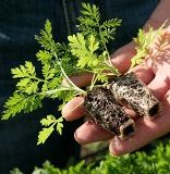
 Artemisinin is the active compound of the simple plant, Artemisia that grows in Southeast Asia. It has been used for years to treat intestinal parasites. The World Health Organization lauds it as a safe malaria treatment. Cancer cells need irons to enable them to grow aggressively; hence cancer cells typically absorb a significantly larger amount of iron than noraml, healthy cells. When Artemisinin come in contact with these irons in the cancer cells, it would trigger a chemical reaction and stimulate increased generation of reactive oxygen species (ROS) and induce apoptosis. The free radicals attack the cancer cell membranes, breaking them apart and killing them. This is why Artemisinin is highly toxic to cancer cells. Tests have been conducted to show that Artemisinin causes rapid and extensive damage and death in cancer cells and yet has relatively low toxicity to normal cells. Unfortunately dietary Artemisinin in supplement form didn’t help cancers due to low solubility and bioavailability. It is recognized that the therapeutic effectiveness of Artemisinin is limited due to its poor absorption from the GI tract, so the use of the natural fermentation to enhance its uptake is particularly beneficial. Artezym is enzymatic fermented Artemisinin. It has perfect bioavailability and pharmacokinetics of active Artemisinin. Artezym is the one and only product that has perfect bioavailability of Artemisinin enough to induce apoptosis in colon cancer cells. Otherwise, Artemisinin wouldn’t have worked.
Heme mediates cytotoxicity from artemisinin and serves as a general anti-proliferation target.
Salts and esters of butyric acid are known as butyrates. Butyrate is an important short chain fatty acid that provides fuel for colon cells and may help protect against colon cancer. The most potent dietary source is butter (3%). The membrane of the GI tract undergoes a complete turnover about every three days. Butyric acid is an important inducer of maturation or differetiation of newly growing intestinal cells. Interestingly, so does vitamin D3. Butyrate might be very useful in the treatment of colon, GIST or stomach cancers. Butyrate is a powerful histone deacetylase (HDAC) inhibitor. HDAC is overexpressed in approximately half of all colon cancer. HDAC inhibitors such as butyrate clearly inhibit the growth of colon cancer cells. Scientists have found a human gene that stops the growth of cancer cells when activated by fiber processing in the colon. Although scientists have long linked butyrate to overall reductions in the incidence of colon cancer, the molecular basis of that benefit has remained largely unknown. Butyrate effects a chemical “unloosening” of molecules that otherwise bind and constrict the activity of the p21 gene. This gene is responsible for the manufacture of p21protein, a compound that slows the growth of cancer cells.
HDAC4 promotes growth of colon cancer cells via repression of p21.
Butyrate inhibits colon carcinoma cell growth through two distinct pathways.
p21(WAF1) is required for butyrate-mediated growth inhibition of human colon cancer cells.
 Butyrate, a fatty acid, comes from two dietary sources. First, it is one of the metabolic end products of unabsorbed dietary carbohydrate that has been bacterially fermented in the gut. Butyrate is the single biggest metabolite of fiber. Second, the only direct source in the diet is from butter, which contains 3% butyrate. Adequate amounts of butyrate are necessary for the health of the large intestine cells. Using good ole butter as a concentrated source of butyrate, we can deliver this natural medicine to the cells of the stomach and GI tract. Vitamin D3 is also a differentiation factor for intestinal cells. Vitamin D3 stimulates the synthesis of calcium binding proteins which promote calcium uptake into the blood. As with butyrate, it also stimulates the maturation of growing intestinal cells. And, of course, it is an excellent natural medicine for the treatment of colon cancer.
Butyrate induces the enzyme that converts inactive vitamin D3 to its active form and stimulates the synthesis of the vitamin D3 receptor (VDR). Butyrate and vitamin D3 synergize to induce p21Waf1, a cell cycle inhibitor that is induced by all HDAC (histone deacetylase) inhibitors. In addition to a lack of inactive vitamin D3 in our bodies due to poor sun exposure, the insensitivity to vitamin D3 found in most cancer cells may be due to enhanced HDAC activity. It is clear that HDAC inhibitors increase vitamin D3 sensitivity in cancer cells. Naturally this translates to enhanced apoptosis in a wide diversity of cancer cells. Therefore, butyrate, like vitamin D3, is a physiological agent that provides an extrinsic signal important in establishing and maintaining colonic mucosal homeostasis. Butyrate decreases cyclin D1 and c-myc expression, each essential for intestinal tumor development, by transcriptional attenuation. Thus, transcriptional attenuation may be a common mechanism involved in normal, well integrated, finely balanced programs of cell maturation in both embryogenesis and in response to physiological inducers of cell maturation in the adult, such as butyrate and vitamin D3 in the intestine.
Transcriptional attenuation in colon carcinoma cells in response to butyrate.
Nutrients regulate the colonic vitamin D system in mice: relevance for human colon malignancy.
Colon-specific regulation of vitamin D hydroxylases–a possible approach for tumor prevention.
Upregulation of 25-hydroxyvitamin D(3)-1(alpha)-hydroxylase by butyrate in Caco-2 cells.
The Vitamin D endocrine system of the gut–its possible role in colorectal cancer prevention.
Vitamin D and colon carcinogenesis.
Short-chain fatty acids and colon cancer cells: the vitamin D receptor–butyrate connection.
Butyrate-induced differentiation of Caco-2 cells is mediated by vitamin D receptor.
1,25-Dihydroxycholecalciferol enhances butyrate-induced p21(Waf1/Cip1) expression.