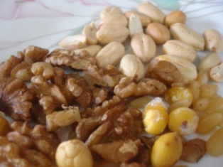
 Phytosterols are plant-derived compounds structurally related to mammalian cell-derived cholesterol that function as essential constituents of plant cell membranes. Phytosterols are composed of 90% sterols and 10% stanols – and scientists maintain the plant ingredients lower cholesterol absorption in the small intestine by competing with it. Both classes of phytosterol are naturally occurring and can be found in a variety of medicinal plants and foods, including nuts, vegetable oils, seeds, and cereals.
Phytosterols act through multiple mechanisms of action, including inhibition of carcinogen production, cancer-cell growth, angiogenesis, invasion and metastasis, and through the promotion of apoptosis of cancer cells. In addition to altering cell-membrane structure and function, phytosterols promote apoptosis by lowering blood cholesterol levels.
Anticancer effects of phytosterols.
New insights into the molecular actions of plant sterols and stanols in cholesterol metabolism.
Phytosterols as anticancer compounds.
Caveolin-1 (Cav-1) is a membrane-anchored protein enriched on caveolae, inverted flask-shaped invaginations on plasma membranes where signal transduction molecules are concentrated. Caveolin-1 (Cav-1) is the main component of caveolae membrane structures and has different roles during tumorigenesis in different cancer types with varying expression profiles, indicating that the role of caveolin-1 varies according to tumor type.
It is highly expressed and acts as a promoter in several tumours like thyroid, prostate, bladder, liver, lung (NSCLC) and pancreatic cancers and in aggressive breast cancers where it is associated with high risk of metastasis and apoptosis suppression. However, study shows that this protein acts as an oncosuppressor in some tumours like not aggressive breast and ovary carcinomas, sarcomas and leukemia.
The Caveolin-1 (Cav-1) and fatty acid synthase (FAS) are two proteins that have been shown to be dysregulated in a number of cancers. Functionally these proteins have been shown to be involved in the process of cancer. Overexpression of these proteins promotes the growth and anti-apoptosis ability of cancer cells. In addition, Cav-1 and FAS overexpression significantly correlated with drug resistance.
The role of caveolin-1 in prostate cancer: clinical implications.
Caveats of caveolin-1 in cancer progression.
Caveolin-1: a tumor-promoting role in human cancer.
As we previously discussed, Fatty acid synthase (FAS) is the major enzyme required to convert carbohydrates to fatty acids. Recent evidence suggests that FAS activity is essential for cancer growth and survival, since blocking the enzyme activity results in cell death. Caveolin-1 (Cav-1) expression is normally required for the upregulation of FAS during cancer progression. Phytosterols regulate expression of Caveolin-1 (Cav-1) and Fatty acid synthase (FAS).
Unlike normal cells, cancer cells lose their ability to respond to death signals that initiate apoptosis (programmed cell death). Sitosterol, the most common plant sterol, has been found to induce apoptosis when added to cultured human prostate, breast, and colon cancer cells.
Caveolin-1 interacts with a lipid raft-associated population of fatty acid synthase.
Cholesterol is an important structural component of mammalian cell membranes. Displacement of cholesterol with phytosterols has been found to alter the physical properties of cell membranes, which could potentially affect signal transduction or membrane-bound enzyme activity. Phytosterols may be linked to increased activity of caspase enzymes. This is achieved by the sterols being incorporated into the cell membranes, resulting in changes to the structure and function of the membranes. These changes ultimately result in an activation of caspase enzymes.
Limited evidence from an animal model of hemorrhagic stroke suggested that very high intakes of plant sterols or stanols displaced cholesterol in red blood cell membranes, resulting in decreased deformability and potentially increased fragility. However, daily phytosterol supplementation (1 g/1,000 kcal) for four weeks did not alter red blood cell fragility in humans.
Considerable emerging evidence supports the inhibitory actions of phytosterols on liver, lung, stomach, colon as well as breast cancer by altering the physical properties of cell membranes. Hence, phytosterols could be incorporated in diet not only to lower the cardiovascular disease risk, but also to potentially kill cancer cells such as thyroid, prostate, bladder, liver, lung (NSCLC), stomach and pancreatic cancers and in aggressive breast cancers.
What’s so special about cholesterol?
beta-Sitosterol inhibits HT-29 human colon cancer cell growth and alters membrane lipids.
Fatty acid desaturase activities are modulated by phytosterol incorporation in microsomes.
Phytosterols are essential for modulating (balancing) the immune system, enhancing it, if it is under active, and reducing it when it is over stimulated. Phytosterols also increase the number and action of natural killer (NK) cells (our cancer fighters) by inducing a Th1, also known as cell-mediated immunity, shift in humans.The effector cells of the TH1 system are cytotoxic T cells and Natural Killer (NK) cells. And increase our DHEA levels naturally. They are also able to reduce the stress hormone cortisol and the proinflammatory immune factor, interleukin-6 (IL-6), and tumor necrosis factor alpha (TNF-a). Interleukin-6 and TNF-a are increased in autoimmune disorders, osteoporosis, over exercising, fibromyalgia, and osteoarthritis. Reduction of this inflammatory agent is the key to halting symptoms and pain. This is exactly what plant sterols and sterolins do.
Chemoprevention of tumor metastasis by liposomal beta-sitosterol intake.
SinnolZym contains powerful anti-cancer phytosterols such as Cerulin and Zorvan extracted from established medicinal plants with a high content of plant sterols. Meanwhile, Capsaicin, the heat in peppers, enhanced the cancer killing properties of SinnolZym. Capsaicin creates a pain response by activating a special group of membrane proteins called vanilloid receptors. When activated, these receptors promote the release of a protein called substance P or SP. It is SP that actually causes pain and inflammation.
Topical capsaicin. The fire of a ‘hot’ medicine is reignited.
tNOX (ENOX2) allows cells to expand in size, which is necessary for proper cellular growth and development. If tNOX is inhibited, the cancer cells die within days. The fact alone could make plasma membrane tNOX the most important anti-cancer target in the cell. Further, it would also be the easiest to inhibit since it is sitting there exposed on the outer cellular membrane. Capsaicin is a tNOX inhibitor. The tNOX inhibitors do not have to enter the cancer cells in order to kill them. Inhibition of tNOX activity by capsaicin correlates with a reduction in growth of cancer cells, indicating a close relationship between tNOX activity and cell growth.
Effect of Ccapsaicin on tNOX (ENOX2) protein expression in stomach cancer cells.
Capsaicin inhibits preferentially the NADH oxidase and growth of transformed cells in culture.
 Capsaicin is a CoQ10 mimic. It has a structure similar to CoQ10 which allows it to compete with CoQ10 in the plasma membrane, mitochondria and lysosome. This is why CoQ10 supplements should not be taken during the cytotoxic phase of a cancer/leukemia treatment protocol. The inhibition of CoQ10 synthesis by statin anti-cholesterol drugs is probably why these drugs are good anti-cancer agents.
Coenzyme Q10, plasma membrane oxidase and growth control.
In the plasma membrane, CoQ10 allows cells to expand in size. It also blocks ceramide release from sphingomyelin while activating sphingosine kinase activity. Sphingosine kinase makes sphingosine-1 phosphate from sphingosine, a breakdown product of ceramide. Spingosine-1 phosphate inhibits cellular death and is a growth factor when secreted into the extracellular environment. In the mitochondria, CoQ10 maintains the electron flow in the inner membrane. If this electron flow is disrupted, cytochrome C will be dislodged from the membrane and enter the cytoplasm. This initiates apoptosis, programmed cell death. Finally, we know that CoQ10 maintains the integrity of lysosomal membranes. If disrupted, the lysosomal interior will cease to become acid, and the lysosomes will leak their contents of iron and cathepsin enzymes into the cytoplasm. This initiates another form of programmed cell death. We believe capsaicin’s ability to compete with and inhibit CoQ10’s activity is the reason it is such a toxic anti-cancer agent.
The following articles suggest other pathways by which capsaicin may initiate programmed cell death.
Capsaicin: a novel chemopreventive molecule and its underlying molecular mechanisms of action.
Immunotherapy of tumors with neuroimmune ligand capsaicin.
Involvement of peroxynitrite in capsaicin-induced apoptosis of C6 glioma cells.
More than spice: capsaicin in hot chili peppers makes tumor cells commit suicide.
Capsaicin probably kills cancer cells in many different ways. SinnolZym and Capsaicin synergistically kill cancer cells. It works. There has been great success in treating many patients with end stage stomach and lung cancer. We will prove it to you in time.

